Abstract
Background: The purpose of this study is to present the X-axis posture improvements in the spine in a young male with adolescent idiopathic scoliosis with occasional complaints of low back pain who was treated with posture correction.
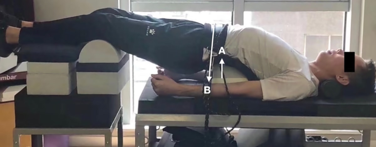
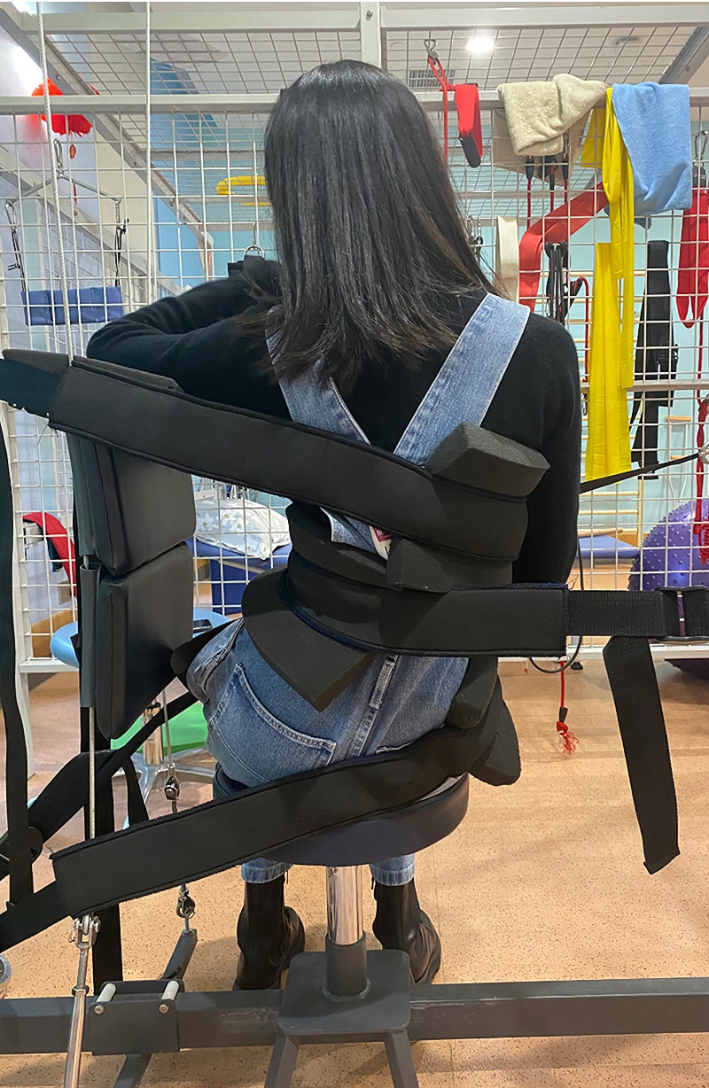
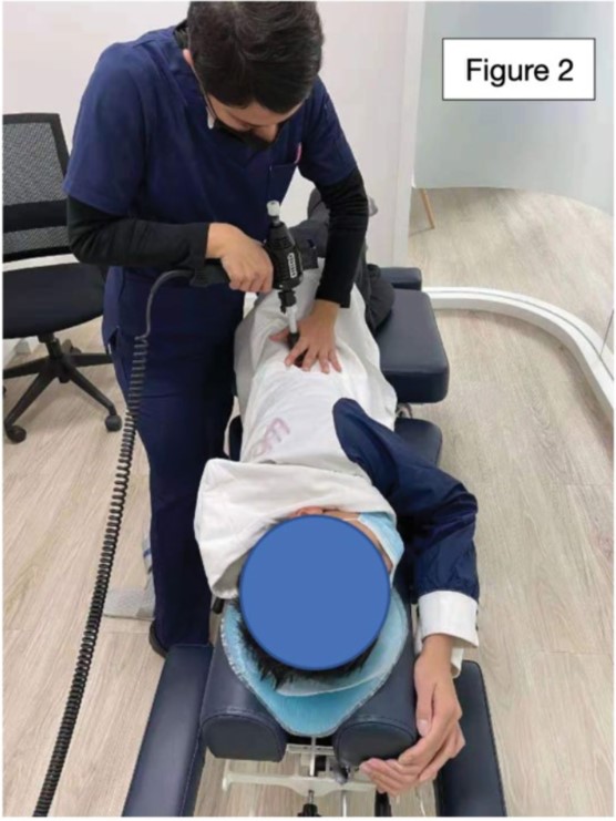
Case presentation: A 15-year-old male presented to the clinic with occasional low back pain. They brought full spine radiographs, (AP and lateral view). Radiographs revealed a cervical kyphosis, forward head, left head tilt, straightening of thoracic kyphosis and lumbar lordosis, and a Cobb angle of 29o with left convexity in the lumbar spine. The patient was treated with ASPINE Systems®; integrating spinal manipulation, corrective exercises and spinal multidimensional traction.
Results: Re-evaluation after 60 sessions during 20 weeks showed improvements in radiographical assessments. The head tilt angle disappeared, deviation of the lower rib cage was aligned to the body’s midline and there was a reduction of the Cobb angle by 17o degrees.
Conclusion: A sizable study using more cases utilizing these protocols and procudures should be conducted to create greater medical awareness of more scoliosis treatment options.
Keywords: ASPINE systems • Adolescent idiopathic scoliosis • Cervical kyphosis • Symptomatic
Introduction
Adolescent Idiopathic Scoliosis (AIS) is a spinal condition causing deformity in 3 dimensions: the coronal, sagittal, and axial planes. AIS is defined as any curve equal to or greater than 10° in the coronal plane [1]. An exact cause of AIS is still unknwon and the etiology includes several factors such as: genetic, hormonal, bone, connective tissue anomalies, biomechanical and neurological factors [2]. AIS has an overall prevalence of 0.47-5.2% with a female to male ratio from 1.5:3.1 and increases substantially with the age [3]. Side curvatures greater than 30° affects 10 times more females than males according to screenings made in elementary and high schools [4].
Treatment for AIS is based on age, curve magnitude and risk of progression that includes observation, physical rehabilitation, orthotic management and surgical correction with spinal fusion; however, experts have recognized that traditional bracing lacks the ability to make 3D corrections, reduces physiological curves, and produces poor cosmetic changes [5]. Surgery is indicated when a curve is progressive despite bracing and when the curve reaches 45o to 50o [2].
Therapists have searched for new treatment methods that can help reduce side curvatures. The Society of Scoliosis Orthopedic Rehabilitation and Treatment (SOSORT) Committee align their guidelines with new evidence based rehabilation procedures and offer recommendations to ensure the clinical practice of Conservative Treatment of Idiopathic Scoliosis (CTIS) is up to date [6].
The International Society of Biomechanics (ISB) suggests human kinematics to be quantified by means of an XYZ Cardan sequence of rotations and translations [7]. The axis represents the body planes, specifically, the three-dimensional view of the spine. Scoliosis due to its nature has postural distortions in multiple planes. X-axis represents the transverse plane, Y-axis represents the frontal plane, and Z-axis represents the sagittal plane. Rotation refers to the angular movement in an axis, for example Rz refers to rotation on the Z-axis. Translation refers to a straight-line movement, such as Tx refers to a translation in the X-axis [8]. XYZ Cardan sequence of rotations and translations was used to analyzed detailed postural shifts and provide correction according to the opposite posture of the scoliotic patient in current case.
Case Presentation
On October 10, 2020, a 15-year-old male came to the clinic presenting postural distortions and previously diagnosed with lumbar scoliosis, suffering from mild low back pain after long periods in the prone position with a Visual Analogue Scale (VAS) 3/10, no other complaints were reported. The teenager underwent rehabilitation for 1 year with no notable changes. The patient reported, “I feel like my left side of the low back is very tight and my right is soft. I did therapy before but I do not feel like there was improvement”. His previous rehabilation program consisted of bilateral thoracic mobility in quadruped position (20 repetitions each side), squats (30 repetitions) and situps (30 repetitions) for a total of 45 minutes twice a week.
Physical examination revealed a decrease lumbar ROM (Range Of Motion) in left lateral flexion and right translation, every other ROM was with in normal limits. Adam’s forward bend test (this test is a simple and non-invasive screening method that requires from the patient to stand up straight with arms at their sides, then bend forward at a 90o degree angle with their arms hanging down as if they are trying to touch their toes) revelead a left lumbar hump with a scale of 16o measured by scolimeter (a type of protactor used to measure the vertebral rotation and rib humping that is seen is scoliosis with forward- bending test).
The patient had several positive orthopedic tests such as cervical distraction and shoulder depression producing pain due to his hypertonic trapezius and scalene muscles. Palpatory findings revealed tenderness and stiffness of the paraspinal muscles in the left side of the lumbar spine due to the severe rotation of the vertebral bodies (confirmed on x-ray). Several spinal fixations were identified at C2, C5, T5-T8 and L4 through static and dynamic palpation. No other orthopedic or neurological test was found positive. Several postural distortions were identified; head tilted to the left from a vertical line, lower left shoulder and lower right iliac crest (assest visually) . On the side view the head, shoulder, hip/pelvis and knee were displaced forward from the vertical line.
The patient brought full spine x-rays (Figure 1) to be assessed via the digitizing image data collection and radiographical interpretation software designed by ASPINE Systems. ASPINE Systems refers to a standard protocol for both spinal diagnostic procedures and noninvasive corrective rehabilitation treatments. The x-ray findings in the front view were: head tilted to the left from vertical line drawn from the middle of the vertebral body of T2 to the middle of the body of C2 creating a tilted angle of 5o degrees. The lumbar spine was shifted 4 cm to the left from center [9]. Cobb Angle gave a measurement of 29o degrees with a left convexity (using lower endplate of L4 and upper plate of T10). Modified Risser Ferguson measurement was 27o degrees using apex mid points of the vertebral bodies of L4, L1 and T10 [10,11].
Coronal view findings were the following: head shifted 2 cm forward from a vertical line starting on the posterior-inferior aspect of C7 and continue all the way up to the posterior aspect of C2; from Harrison Posterior Tanget method a cervical kyphosis was visible at C5-C7 with Hypo-lordosis measurement of -8o (normal value of -42o) and Hypo-kyphosis on the thoracic spine with a measurement of +22o (normal value of +37o) [7].

blog-4-1
Figure 1. Lumbar X-ray viewed posterior to anterior. Cobb angle of 29o/ Risser Ferguson of 27o; left translation (+TxT) by 4 cm from center.
Methodology
The patient consented to posture corrective treatment targeting the both Z-axis and X-axis posture diviations. The patient agreed to a 3 session per week treatment plan; each session time lasted 60 minutes to 90 minutes.
Each session the patient was checked for spinal fixations with dynamic palpation and was corrected using a Chiropractic drop table. Posture correction was perform on the side lying position requiring the patient to perform a mirror- image position while a percussion device was utilized to produce a succession of rapidly repeating, high velocity-low amplitude thrusts to activate the patient’s proprioceptors and mechanoreceptors (Figure 2).

blog-4-2
Figure 2. High velocity-low amplitude thrusts to activate the patient’s proprioceptors and mechanoreceptors.


